Keratoconus

Keratoconus is a relatively common condition, affecting 1 in 1800 people. This condition causes the cornea to become cone shaped as the result of a bulging weak spot.

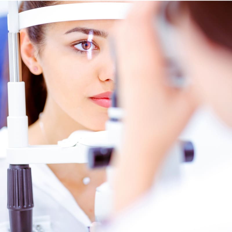
brown-eyed-woman-with-light-beam-on-her-eye-in-ophtalmologists
Centre for Sight - Keratoconus - eye
Consultants at Centre for Sight working together

What causes Keratoconus?

Keratoconus is a condition which causes the cornea to become cone shaped as the result of a bulging weak spot. This bulge creates distortion in your vision, so those with the condition have difficulty focussing. Keratoconus is more likely to progress in those who develop the condition at an early age. It involves both eyes, but can affect one eye more than the other due to development of the condition at different rates. Eye rubbings worsens the condition and it is often associated with allergies, asthma and eczema.

Marcela Espinosa-Lagana
Cornea, Cataract, Keratorefractive Surgery and Paediatric Ophthalmology

There is no single cause for the condition Keratoconus and numerous factors have been implicated and the causes are “multifactorial”. Many with Keratoconus are concerned that they may pass on the condition to their children. Whilst Keratoconus can be inherited, a more common association is allergies and patients often have hay fever, asthma and eczema. Hay fever causes itchiness and eye rubbing aggravates the condition.

Although Keratoconus can occur earlier, more often its onset is in puberty. The condition progresses and then in the late 30s and 40s slows down and stops. Keratoconus has also been observed in contact lens wearers and it is not clear if contact lenses can contribute to the condition or is it because Keratoconus results in shortsightedness and patients use contact lenses for correction. Overall in Keratoconus, the cornea is more elastic than normal and has a tendency to change shape and become steep. Pressure on the cornea can make the weakest area bulge more and is the reason why eye rubbing is strongly discouraged.

Read More

Treating Keratoconus

Tired asian business woman with headache at office, f
Spectacles/Glasses

In the early stages when the cornea is not too abnormally shaped, glasses can help correct vision. Often glasses need to be changed frequently and this is the tip-off that the patient may have Keratoconus, especially if there is a progressive increase in astigmatism.

brown-eyed-woman-with-light-beam-on-her-eye-in-ophtalmologists
Hard Contact Lenses

Hard or rigid contact lenses and Scleral lenses are useful for improving vision when glasses no longer work. A hard contact lens evens out the corneal irregularities and permits good vision. In some cases contact lenses can become difficult to fit or tolerate at which point surgical options may need to be considered.

Centre for Sight patient during a consultation
CXL – Corneal Collagen Cross linking with Riboflavin

This procedure is used to strengthen the cornea and has been used at Centre for Sight since 2005. Strengthening the cornea and reducing its elasticity can stop progression and in some case even cause flattening and an improvement in corneal shape. Find out more about CXL by clicking here.

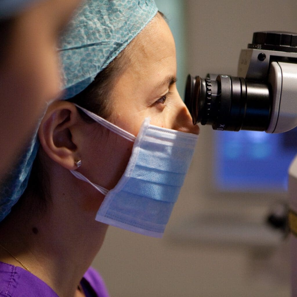
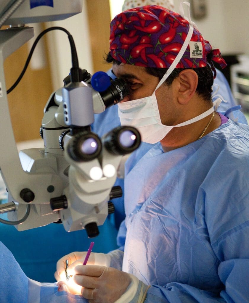
Centre for Sight surgeon Marcela Espinosa-Lagana using state of the art technology at the clinic
Keranatural CAIRS

The use of corneal tissue to alter the shape of the cornea is a recent advance in the treatment of keratoconus. Results of this procedure have been outstanding and now the preferred option for Intracorneal ring segments. Plastic inserts (Intacs and Ferrara – right) have been used with great success in the past, however the use of cornea avoids halos and light reflection. Learn more about Keranatural CAIRS here.

Catract-patient-at-Centre-for-Sight-with-consultant-Marcela-Espinosa-
Intracorneal Rings (Intacs and Ferrara Rings)

These are rings that are inserted into the cornea using a femtosecond laser to create channels. The rings alter the shape of the cornea regularising the shape decreasing astigmatism as well as reducing shortsightedness / myopia by flattening the central cornea. For those who have become intolerant to contact lenses and where the cone is not scarred or not excessively advanced, intracorneal rings can help improve vision with glasses or even allow contact lenses to be fitted. For those who obtain improved vision with glasses, Toric Implantable Contact lenses can be considered to eliminate the need for optical aids altogether. Learn more about Intracorneal Rings here.

Centre for Sight leading consultant and Medical Director Sheraz Daya
Toric Implantable Contact Lenses

For those who are able to obtain good vision with glasses and have Keratoconus that has stopped progressing, Toric Implantable Contact Lenses can reduce the need for glasses and in many cases eliminate the need for optical aids. Implanting Toric ICLs in patients with Keraotoconus is less predictable than regular myopic astigmatis. You can learn more about the Toric ICL here.

Femtosecond Laser PK
Corneal Transplants

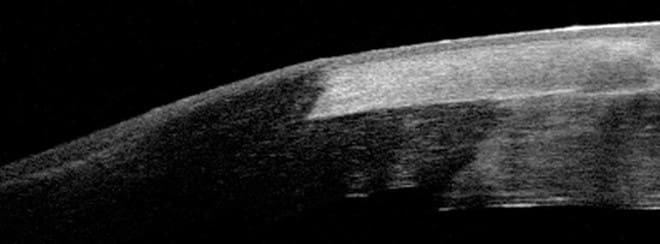
In very advanced stages affecting approximately 15% of Keratoconus patients a corneal transplant may be needed. A partial thickness or Deep Anterior Lamellar (DALK) is the preferred option. A full thickness or Penetrating keratoplasty (PK) is an option that has been used in the past and in countries outside the USA is becoming less common. Precise lasers for use in laser vision correction have been adapted to enable grafting procedures. This technique improves corneal graft apposition and mechanical strength. The option of a Femtosecond Laser Deep Anterior Lamellar Keratoplasty has been developed and pioneered by Centre for Sight surgeons. The technique displayed in a video created by Centre for Sight was the recipient of a video award at the American Society of Cataract and Refractive Surgeons, San Francisco, 2013. You can learn more about Corneal transplants here.

Surgeons at Centre for Sight are internationally renowned for their expertise in Keratoconus and Corneal problems. Centre for Sight’s medical director is on numerous boards and committees related to Keratoconus and is on the founding editorial board of the peer-reviewed publication Keratoconus, as well as a founding member of the Keratoconus Expert Group, an international group of Keratoconus specialists. He was involved as a panellist in the Global Consensus Working Group the findings and conclusion of which were recently published in the journal Cornea.

Author Information

Authored by Sheraz Daya MD FACP FACS FRCS(Ed) FRCOphth, Consultant Ophthalmic Surgeon & Medical Director, June 2019.

Next review due June 2025.